Day 2 :

Biography:
Huang Wei Ling, born in Taiwan, raised and graduated in medicine in Brazil, specialist in infectious and parasitic diseases, a General Practitioner and Parenteral and Enteral Medical Nutrition Therapist. Once in charge of the Hospital Infection Control Service of the City of Franca’s General Hospital, she was responsible for the control of all prescribed antimicrobial medication and received an award for the best paper presented at the Brazilian Hospital Infection Control Congress in 1998. Since 1997, she works with the approach and treatment of all chronic diseases in a holistic way, with treatment guided through the teachings of Traditional Chinese Medicine and Hippocrates. Researcher in the University of São Paulo, in the Ophthalmology department from 2012 to 2013.Author of the theory Constitutional Homeopathy of the Five Elements Based on Traditional Chinese Medicine. Author of more than 100 publications about treatment of variety of diseases rebalancing the internal energy using Hippocrates thoughts.
Abstract:
Keynote Forum
Muhammad Mumtaz
King Salman Hospital, KSA
Keynote: Can there be a correlation established between characteristic histological and radiographical findings in adenomatoid odontogenic tumor? A case report
Time : 10:20-11:00

Biography:
Abstract:
Keynote Forum
Lincoln Harris
University of Queensland, Australia
Keynote: The Art of Treatment Planning
Time : 09:00 - 09:40

Biography:
Abstract:
- 3-D imaging in dentistry, Clinical and Medical case reports, Conservative Dentistry, Craniofacial Surgery, Dental Biomaterials & Bioengineering, Dental Caries, Dental Education, Dental Implants, Dental Marketing
Location: Hall 1
Session Introduction
Jennifer M de St Georges
JdSG International Inc
Title: A Dentist is Judged by Everything but Their Quality of Care’ ...3 Steps to elevate your practice/patient communication & enjoy increased patient demand and treatment acceptance
Time : 12:10-12:40
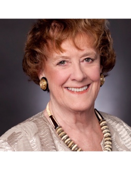
Biography:
Mrs. de St. Georges has presented more than 1,000 full day dental practice management programs on 6 continents. She has presented for eight FDI Conferences. She has published more than 300 articles in US and international journals. Ms. de St. Georges was the first non-dentist to be appointed by USA’s leading journal Dentistry Today to their Editorial Board. Born and raised in the UK, she has been based in Northern California, USA., for many years.
Abstract:
Overview: Dentists do not sell dentistry. Patients ‘buy’ your services. When patients are educated to appreciate their treatment benefits, their financial investment becomes a non-issue. Your new problem? ...finding open time in your schedule!
---a referred patient has ‘bought’ before walking through your door
---money is never an issue when patients perceive the value to be enjoyed
---how ‘inform before you before’ must become your practice core message & management style
---patients attracted by convenience, will leave when it becomes inconvenient-to them
---understand how today’s technical marketing tools have potential of sabotaging your reputation
---learn how to discuss and collect fees while feeling cool, calm, confident and comfortable
Majority of diagnosed dentistry is elective-moving patients from ‘let me think about it’ to ‘YES’
Outcome: Attendees now have tools to evaluate the effectiveness of their current management approach and adjust accordingly to encourage patient commitment and practice growth
Mohamed Abdulcader Riyaz
Qassim University, KSA
Title: Current trends in Salivary Gland Imaging – A Review
Time : 12:40-13:10

Biography:
Abstract:
Mohammed Dubais
University of Science and Technology College of Dentistry, Yemen
Title: The effect of crude Khat extract on the color change of Nano Composite Resin material

Biography:
Mohammed Ahmed Dubais has completed his Bachelor’s degree from King Saud University, Saudi Arabia. He has completed his PhD from University of Khartoum Sudan. He was the Vice Dean, College of Dentistry, UST and Lecturer of Prosthodontics. He was also the Member of the Editorial Board of the Yemeni Dental Journal. He has served as a Dean at College of Dentistry UST till 2011.
Abstract:
The use of resin composite for restoring teeth has been increased greatly in recent years; manufacturers are claiming that the nanofilled composite technology has been improved to be similar to the ceramics in shade selection and color stability. In Yemen, khat chewing habits may affect the color of esthetic restoration and it is always accompanied by drinking water and cola.
- Dental Marketing, Dental Material Sciences ,Dental Nursing Dental Nursing and Public Health Dentistry ,Endodontics, Forensic Odontology ,Holistic Dentistry, Maxillofacial Pathology, Microbiology & Surgery, Mouth Cancer Oral Cancer Research Orthodontics Paediatric Dentistry Periodontology and Implant Dentistry, Public Health Dentistry
Location: Hall 2
Session Introduction
Zainal Arif Abdul Rahman
University of Malaya, Malaysia
Title: Orofacial Vascular Lesions
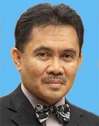
Biography:
Zainal A A Rahman is presently working as Dean at Faculty of Dentistry, University of Malaya, Malaysia. Currently, he is working as a Professor at Department of Oro-Maxillofacial Surgical & Medical Sciences, Faculty of Dentistry, University of Malaya, Malaysia. He has published more than 38 papers in reputed journals.
Abstract:
Vascular lesion within the mouth, face and jaws had pose an unsolved challenge to the oral and maxillofacial surgeons due to possible risk of excessive blood loss during surgical intervention and also frequent recurrence of the lesion. The vascular lesions that occur commonly within orofacial region include arteriovenous malformation, cavernous haemangioma and vascular tumor. The author will describe the diagnosis and various treatment options for the variety of vascular lesions. The rational and justification of each treatment option will be discussed in detail. The need of prior embolization in relation to the type of lesion will also be emphasized. The merit and disadvantage of each treatment options will be discussed over a range reported cases. The complications that may arise from these lesions will also be mentioned.
Sinem Yeniyol
Istanbul University, Turkey
Title: Non-UV Based Early Murine Pre-Osteoblastic Cell ALP Levels of Anodized and Annealed Titanium Surfaces

Biography:
Abstract:
Abhijeet Bhasin
World Academy of Ultrasonic Piezoelectric Surgery, India
Title: Immediate Dental Implant Placement in Extraction Socket
Biography:
Abhijeet Bhasin has completed his graduation from Modern Dental College and Research Centre, Indore in 2007 and has been practicing Implantology since then. He has done his Basic and Advanced Implantology training from Goethe University, Germany under the mentorship of Dr. Prof. Netwig and Dr. Porus Turner. He is a Member of Indian Society of Oral Implantologists (ISOI) and Vice President of Indian Dental Association, Indore Branch. He is the Vice President of World Academy of Ultrasonic Piezoelectric Surgery (WAUPS) South East Asia and the Fellow of Academy of General Education and World Academy of Ultrasonic Piezoelectric Surgery.
Abstract:
Immediate dental implant placement has been an acceptable procedure from at least the past two decades. Commonly, immediate implants have been reserved for the single rooted anterior tooth and single or bi-rooted premolar tooth. Perhaps the most important aspect of any implant surgery in accordance with the successful procedure is implant stability and bone to implant contact (BIC). Removal of molar teeth provides a challenging and intriguing dilemma due to multiple root morphology. In the case of extraction and immediate placement of dental implants preserving alveolar bone proper, particularly that of the labial and lingual plates of bone is essential in providing the optimal environment for maximizing BIC and implant stability. Also, the position of the final restoration must be considered, in relation to intra and inter arch position, occlusion, function and aesthetics. Thus, minimal alveolar bone removal should be considered and attained to aid in the above factors in order to provide an acceptable surgical site for successful placement of the dental implant. Finally, and perhaps most importantly when considering immediate molar implant placement, removal of the intra-alveolar septum should be avoided to aid in increasing BIC and allowing the attainment of initial implant stability at the time of placement.
